Eye Care > Diseases of the Eye > Retinal Vein Occlusion
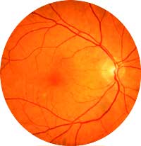
Normal Retina.
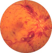
Retinal Vein Occulsion.
Vein occlusion is diagnosed by examining the retina with an ophthalmoscope. Fluorescein angiography may be performed in some cases to study the circulation of the retina and to determine the extent of macular oedema or swelling.
Following a vein occlusion, the primary concern is to treat the secondary complications.If areas of the retina are oxygen-deprived, LASER may be used to prevent growth of delicate vessels that could break, bleed or cause glaucoma.
The following are common risk factors for vein occlusion:
Illustrations by Mark Erickson
With acknowledgement to St. Lukes Eye Hospital.
| Cookie | Duration | Description |
|---|---|---|
| cookielawinfo-checkbox-analytics | 11 months | This cookie is set by GDPR Cookie Consent plugin. The cookie is used to store the user consent for the cookies in the category "Analytics". |
| cookielawinfo-checkbox-functional | 11 months | The cookie is set by GDPR cookie consent to record the user consent for the cookies in the category "Functional". |
| cookielawinfo-checkbox-necessary | 11 months | This cookie is set by GDPR Cookie Consent plugin. The cookies is used to store the user consent for the cookies in the category "Necessary". |
| cookielawinfo-checkbox-others | 11 months | This cookie is set by GDPR Cookie Consent plugin. The cookie is used to store the user consent for the cookies in the category "Other. |
| cookielawinfo-checkbox-performance | 11 months | This cookie is set by GDPR Cookie Consent plugin. The cookie is used to store the user consent for the cookies in the category "Performance". |
| viewed_cookie_policy | 11 months | The cookie is set by the GDPR Cookie Consent plugin and is used to store whether or not user has consented to the use of cookies. It does not store any personal data. |